


Helminthiasis (helminthic infestation): causes, symptoms, diagnosis and treatment methods.
Ohdetermination
Helminthiases are diseases of humans, animals and plants caused by parasitic worms (helminths).
Causes of helminthiasis
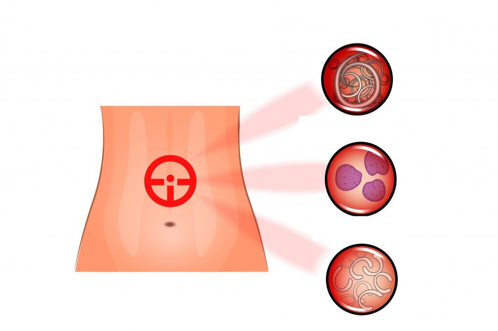
Currently, in our country there are more than 70 species of the 250 known helminths that parasitize the human body.The most common are nematodes (roundworms, pinworms, trichinella, whipworms), tapeworms (pig, bovine and dwarf tapeworms, broad tapeworm, echinococcus) and trematodes (liver and cat flukes).
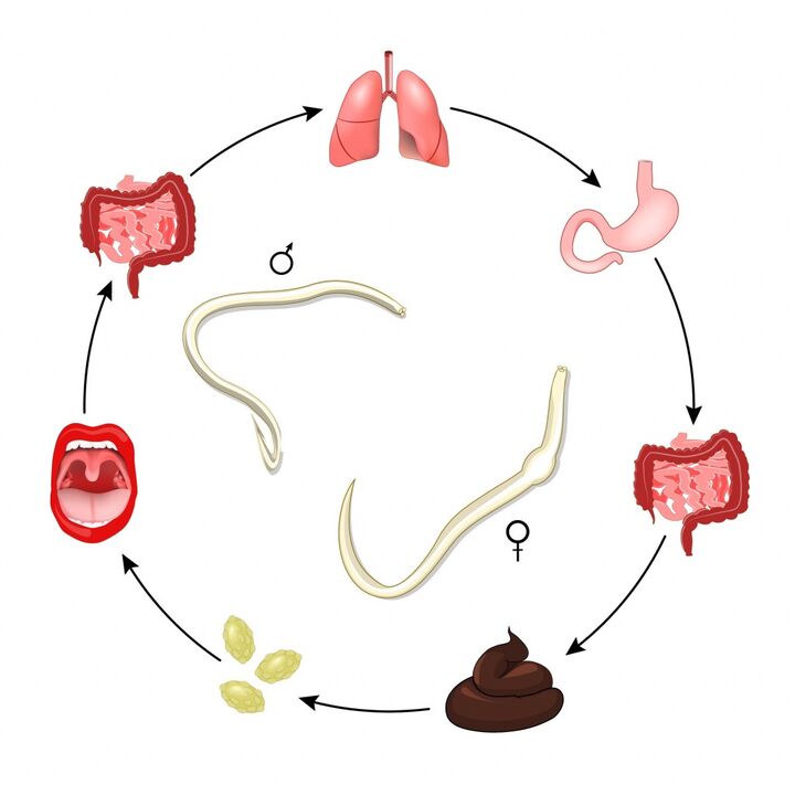
Helminths are characterized by stages of development: eggs → larvae → sexually mature forms.
Helminth infection most often occurs after their eggs and/or larvae enter the body.According to the infection mechanism and transmission routes, helminthiases are divided into: soil-transmitted helminthiases, biohelminthiases and contact helminthiases.Geohelminths develop without intermediate hosts, biohelminths develop with a sequential change of one, two or three hosts, contact helminths are transmitted by contact.
The pig tapeworm, bovine tapeworm, echinococcus and other types of worms develop with a successive change of one, two or three hosts.Intermediate hosts can be fish, mollusks, crustaceans and insects.A person becomes infected with these helminths by eating food that has not undergone complete heat treatment:
- beef infected with bovine tapeworm larvae;
- pork affected by the Finnish pork tapeworm;
- raw and lightly salted fish with opisthorchis or broad tapeworm larvae;
- raw water or vegetables and fruits treated with this water.
Whipworms, roundworms, hookworms, and necators develop without intermediate hosts.The eggs and larvae of these parasites enter the soil with feces.If the rules of personal hygiene are not followed, they penetrate the body of a new host.
By contact, that is, through personal contact between a healthy person and an infected person, through the use of shared utensils, toiletries, bedding and through inhalation of dust in a room where an infected person is located, enterobiasis (pathogen - pinworms) and hymenolepiasis (pathogen - dwarf tapeworm) are transmitted.In the case of enterobiasis, autoinfection often occurs.
Helminths of a certain type parasitize certain organs and cause various helminthiases:
- in the large intestine: pig, bovine, dwarf tapeworms, nematodes (hookworms, roundworms, strongyloids), pinworms, whipworms.From the intestinal lumen, pork tapeworm larvae can enter the bloodstream and spread throughout the body, settling in adipose tissue, muscle vessels, eye chambers and brain;
- in the liver and biliary tract: trematodes (opisthorchis, clonorchis, fasciola).Echinococcal cysts are mainly located in the liver, and after their rupture, daughter blisters can be found in the mesentery, peritoneal layers, spleen and other organs;
- in the respiratory organs: echinococci, alveococci, lung flukes, which cause paragonimiasis;
- in the nervous system: schistosomiasis, paragonimiasis, echinococcosis and alveococcosis;
- in the organs of vision: onchocerciasis, loiasis, complicated forms of taeniasis;
- in the circulatory system: necatoriasis, schistosomiasis, diphyllobothriasis;
- in the lymphatic system - filariasis, trichinosis;
- on the skin and subcutaneous tissue: hookworm, onchocerciasis, loiasis, larval stage of schistosomiasis;
- in the skeletal system - echinococcosis;
- in skeletal muscles: trichinosis, cysticercosis of muscle tissue.
The lifespan of helminths in the organism of the definitive host can be different, depends on the type of parasite and ranges from several weeks (pinworms) to several years (tapeworm) and decades (fasciola).
Classification of the disease.
There are two types of worms that parasitize humans:
- Nemathelminthes – roundworms, class Nematoda;
- Flatworms are flatworms, which include the classes Cestoidea (tapeworms), Trematoda (the class trematodes).
Depending on the routes of spread of the parasites and the characteristics of their biology, they are distinguished:
- biohelminthiasis;
- soil-transmitted helminthiasis;
- Contact helminth infections.
Symptoms of helminthiasis.
Helminths have different effects on the human body:
- antigenic effects, when local and general allergic reactions develop;
- toxic effect (helminth waste products cause malaise, weakness, dyspeptic symptoms);
- traumatic effect (when parasites attach to the intestinal wall, the blood supply is disrupted with necrosis and subsequent atrophy of the mucous membrane; absorption processes may be disturbed; mechanical compression of tissues by helminths);
- secondary inflammation as a result of the penetration of bacteria after the migration of helminth larvae;
- violation of metabolic processes;
- as a result of the absorption of blood by some helminths, anemia occurs;
- neuroreflex effect: irritation of nerve endings by helminths causes bronchospasm, intestinal dysfunction, etc.;
- psychogenic effect, manifested by neurotic conditions, sleep disturbances;
- immunosuppressive effect.
Helminthiases are characterized by stages of development.Each stage is characterized by its own clinical symptoms.
In the initial acute stage, helminths often do not yet release eggs, sensitization of the body occurs (production of antibodies, release of inflammatory mediators, increased permeability of the vascular wall) and traumatization of the organs through which the larvae migrate.Clinical symptoms may be absent, but in some cases the disease may present with pronounced clinical manifestations.The acute stage lasts 1 to 4 months, sometimes 8 to 10 months or more.
Complaints of patients in the acute stage:
- increase in temperature from several days to two months (mild fever or higher than 38ºС, accompanied by chills, severe weakness and sweating);
- recurrent itchy skin rashes;
- local or generalized edema;
- enlargement of regional lymph nodes;
- pain in muscles and joints;
- cough, asthma attacks, chest pain, prolonged cold symptoms, bronchitis, tracheitis, symptoms simulating pneumonia, asthmatic syndrome, hemoptysis;
- abdominal pain, nausea, vomiting, stool disorders.
At the end of this stage of helminth infection, acute allergic phenomena gradually subside, and the clinic of the chronic stage has not yet had time to develop.
The acute stage becomes subacute when the "young" helminths gradually mature.Next comes the chronic stage, corresponding to the development of parasites in sexually mature individuals.The clinical picture develops against the background of the toxic effect of waste products of helminths, the traumatic effect of helminths on organs (hookworm, trichuriasis, etc.), mechanical effects (an echinococcal cyst in the liver grows, compresses neighboring organs; cysticerci - in the brain), a secondary inflammatory process (duodenitis is observed in strongyloidiasis), metabolic disorders (hypo- or avitaminosis),functional disorders of the stomach and duodenum, secondary immunodeficiencies, etc.
The symptoms depend on the organs in which the helminths parasitize, their size and quantity.
Forintestinal helminthiasisThe following syndromes are characteristic:
- dyspeptic (upset stomach, feeling of fullness after eating, early satiety, bloating, nausea);
- painful;
- asthenoneurotic (feeling of extreme fatigue, increased nervous excitability and irritability).
Enterobiasis is characterized by nocturnal perianal itching.With massive roundworm infestation, intestinal obstruction, pancreatitis, and obstructive jaundice may occur.
Intestinal cestodes(taeniarinhoz, diphyllobothriasis, hymenolepiasis, taeniasis and others) are asymptomatic or with a small number of symptoms (with symptoms of dyspepsia, pain, anemia).
Liver flukes(fascioliasis, opisthorchiasis, clonorchiasis) cause:
- chronic pancreatitis;
- hepatitis;
- cholecystocholangitis;
- neurological disorders.
Hookworm diseaseIt is manifested by asthenovegetative syndrome (weakness, fatigue, pale skin) due to the development of iron deficiency anemia.
Filariasis is characterized by an allergic syndrome of varying severity and regional lymphadenitis.
Urogenital schistomiasisIt is manifested by the appearance of blood at the end of urination, frequent need to urinate, pain when urinating.
Alveococcosis, cysticercosis, echinococcosis.can remain asymptomatic for a long time.At a later stage, suppuration or rupture of parasite-containing cysts causes anaphylactic shock, peritonitis, pleurisy and other serious consequences.
For diseases caused by parasitism of migrating larvae.zoohelminthswhen a person is not the natural host, distinguishes between cutaneous and visceral forms.The cutaneous form is caused by the penetration of certain animal helminths under the human skin: schistosomatids of waterfowl (trematodes), hookworms of dogs and cats, strongylids (nematodes).When a person comes into contact with soil or water, helminth larvae penetrate the skin.There is a burning, tingling or itching sensation at the place of introduction of the helminth.There may be a brief fever and signs of general malaise.After 1-2 weeks (less often - 5-6 weeks), recovery occurs.
The visceral form develops as a result of ingestion of helminth eggs with water and food.At the beginning of the disease there may be general malaise, allergic exanthema (skin rash).In the human intestine, larvae hatch from helminth eggs, penetrate through the intestinal wall into the blood, reach the internal organs, where they grow and reach 5-10 cm in diameter, compress the tissues and disrupt the function of the organs.When tapeworm larvae (cysticerci, tsenura) are localized in the membranes and substance of the brain, headaches, signs of cerebral hypertension, paresis and paralysis, and epileptiform seizures are observed.The larvae can also be located in the spinal cord, eyeball, serous membranes, intermuscular connective tissue, etc.
The result of helminthiasis can be a complete recovery with the elimination of helminths or the development of irreversible changes in the host's body.
Diagnosis of helminthiasis.
The diagnosis of helminthiasis is established on the basis of a combination of complaints, information received from the patient about the course of the disease, laboratory data and instrumental examination methods.
In the acute phase of helminth infections, a blood reaction occurs due to the presence of a helminth in the body, which is why the following studies are recommended:
- clinical blood test: general analysis, leucoformula, ESR (with microscopy of a blood smear in the presence of pathological changes);
- biochemical blood test:
- total protein, albumin, protein fractions;
- evaluation of kidney function indicators (urea, creatinine, glomerular filtration);
- evaluation of liver function indicators (bilirubin, ALT, AST, alkaline phosphatase);
- pancreatic alpha-amylase;
- Assessment of carbohydrate metabolism: glucose (blood), glucose tolerance test with determination of fasting and post-exercise venous blood glucose after 2 hours.
The following biological materials can be examined for the presence of helminths: feces, blood, urine, duodenal contents, bile, sputum, muscle tissue, rectal and perianal mucus.
Since helminths are not excreted in feces at any stage of their development, it is recommended to donate feces three times, every 3-4 days.
- Analysis of feces for helminth eggs.
- Enterobiasis (pinworm eggs) test, swab.
- Microscopic examination of feces to detect eggs of the enterobiasis pathogen Enterobius vermicularis (pinworms).
- Test for enterobiasis (pinworm eggs), spatula.
- The analysis is necessary if there is suspicion of pinworm infection, as well as during hospitalization and recording a medical history.
- Analysis for kindergarten and school.
The child screening program must be completed before entering kindergarten or school.The results of these studies are necessary to obtain a medical certificate according to form 026U approved by the Ministry of Health of the Russian Federation.
Perform immunological studies: determination of specific antibodies against helminths:
- IgG anti-intestinal worm antibodies;
- anti-Echinococcus IgG;
- IgG antibodies against Toxocar antigens;
- anti-Opisthorchis felineus IgG (IgG class antibodies against feline fluke antigens);
- IgG antibodies against Trichinella antigens;
- antibodies against the causative agent of anisakiasis (nematodes of the genus Anisakis), IgG;
- antibodies against the causative agent of clonorchiasis, IgG;
- Antibodies against Strongyloides stercoralis, the causative agent of strongyloidiasis, IgG.
A test intended to detect IgG antibodies against the causative agent of strongyloidiasis is used for early serological diagnosis of strongyloidiasis in cases of clinical suspicion (eosinophilia, serpentine skin lesions, pulmonary or gastrointestinal symptoms) of this disease.
In difficult cases, the following studies may be recommended:
- simple x-ray of the chest organs;
- full ultrasound examination of the abdominal organs (liver, gallbladder, pancreas, spleen);
- Computed tomography of the abdominal cavity and retroperitoneum;
- CT scan of the chest and mediastinum;
- CT scan of the brain and skull.
Which doctors should I contact?
If you suspect helminthiasis, you should consult a therapist or family doctor and, with your child, a pediatrician.
If there are indications for surgical treatment, the patient is referred to a surgeon.
Helminthiasis treatment
Most people with helminth infections do not require hospitalization.Patients with tissue helminthiasis, regardless of its severity, and patients with a severe and complicated course of the disease are treated in the hospital.
Therapy is carried out with anthelmintic drugs.The frequency of administration and dosage of the drug depend on the age and body weight of the patient and are established by the doctor.
Desensitizing, detoxifying and vitamin therapies may be indicated.To reduce the temperature, non-steroidal anti-inflammatory drugs are prescribed, for an allergic reaction and itching - antihistamines, for severe edema - diuretics.In severe cases, hormonal medications are required.
The presence of helminths in organs and tissues may be an indication for surgical treatment.
Complications
- bronchial asthma.
- Pneumonia.
- Bowel cancer.
- Cirrhosis.
- Portal hypertension.
- Gastrointestinal bleeding.
- Ascites.
- Hepatitis.
- Liver abscess.
- Peritonitis.
- Allergic myocarditis.
- Meningoencephalitis.
- Chronic glomerulonephritis.
- Chronic kidney disease.
- Hemostasis disorders.
- Loss of vision.
Helminthiasis prevention
Preventive measures aimed at preventing helminth infection include:
- compliance with personal hygiene standards (use of individual towel, personal hygiene items and other accessories for daily use);
- use only high-quality water in everyday life;
- periodic vaccination and deworming of pets;
- Thoroughly washing vegetables, fruits and berries before consumption;
- Sufficient heat treatment of meat and fish products.
IMPORTANT!The information in this section cannot be used for self-diagnosis or self-treatment.In case of pain or other exacerbation of the disease, diagnostic tests should be prescribed only by the treating doctor.To make a diagnosis and properly prescribe treatment, you must contact your doctor.